www.BioTopics.co.uk
Site author Richard Steane
The BioTopics website gives access to interactive resource material, developed to support the learning and teaching of Biology at a variety of levels.

Mass transport in animals

Mouseover of green text (in the main body, not headings) should bring in further explanation in a small popup window.
Clicking a couple of underlined words should result in spoken help in pronunciation.
Let me know if you think I should do this for other words.

Mass transport: a delivery system for lots of molecules

Every single cell of the human body needs a supply of oxygen and glucose, which it uses for (aerobic) respiration. As a result, it produces carbon dioxide and water, which need to be removed.

Although cells take in oxygen and give out carbon dioxide by diffusion, this is a slow process and when cells are combined to form tissues and organs within the body of a multicellular organism, there must be some specialisation to provide what cells need, as well as removing what they do not need.

The circulatory system provides a flow of blood to all organs of the body, leading from respiratory surfaces and the digestive system and taking wastes to excretory organs. The blood is pumped through blood vessels and it acts as a mass transport system for all these substances,

The substances all travel together in the blood at the same speed, although the flow rate may be varied by the pumping mechanism and the dynamics of the blood vessels supplying different organs. At this level, different organs can take in what they need or get rid of what they do not need. These substances are transferred by diffusion.

Blood carries these these substances in solution (in water) but the efficiency of transport and delivery may be improved by other compounds.

Plasma proteins

This topic covers the transport of oxygen and carbon dioxide, so it is a natural follow-on to gas exchange, and it centres on the intracellular protein haemoglobin and blood.

There are other proteins - plasma proteins - in blood, and they have functions in the transport of other substances which are sometimes thought to be simply soluble in water.

Serum albumin accounts for about 55% of blood proteins. It has a large slightly charged molecule and it forms a colloidal solution. It has an osmotic effect but significantly it cannot cross capillary walls. It thus affects the water potential of blood and has a part to play in the movement of tissue fluid out from the blood and back into it. See below
It also functions as a carrier of many components of blood, notably steroidal hormones and other lipids. It is a factor in the delivery of certain drugs and can influence the dose rate.

Globulins make up about 38% of blood proteins. These globular proteins also transport ions, hormones, and lipids. Antibodies - the main tools in the immune system acting against disease organisms - are classed as immunoglobulins.

About 7% of blood protein is fibrinogen. This is involved in blood clotting.

There are also variable amounts of other proteins carried in the blood plasma, such as hormones, enzymes and regulatory proteins.

Haemoglobin

is a compound that increases the oxygen-carrying capacity of blood, compared with water alone.
It gives blood its red colour.

It is a protein with a fairly complex molecular structure. Being composed of 4 polypeptide chains, it has a quaternary structure, and each of these chains is attached to a haem group containing iron. There are in fact two α- and two β- globin chains, and it may be described as a tetramer. Each α chain is built up from 141 amino acid residues and each β chain is 146 amino acid residues long. These chains are coiled, and the structure is stabilised by internal bonds.

A molecule of human haemoglobin haemoglobin (38K)
The polypeptides are coiled into a globular shape, and the haem group is a porphyrin, a fairly flat network or macrocycle of carbon and nitrogen atoms, with a single atom of iron in the centre. This iron atom can form a loose association with molecular oxygen (O2), so a single haemoglobin molecule can carry (or release) up to 4 molecules of oxygen.

There are minor differences in the structure of haemoglobin in different organisms, most of which are due to differences in the amino acid sequence of the polypeptide section of the molecule. This can result in variation in the oxygen-carrying capacity of the molecule which effectively adapts the organism to their environment.

Haemoglobin is a very efficient means of transporting - but not storing - oxygen around the body.

But it also interacts with other compounds in the body - not just gases. See below

There are many forms of haemoglobin

Haemoglobin is found in many groups within the animal kingdom and they use it to pick up and carry oxygen.

Fetal mammals produce haemoglobin with a different structure to enable them to absorb oxygen more efficiently from their mother's blood stream.

Animals inhabiting environments with lower oxygen concentrations have haemoglobin that maximises their uptake of oxygen.

Animals inhabiting higher altitudes have differences in their haemoglobin to enable them to retain as much oxygen as possible in their 'thin' atmosphere.

It is also found (as 'leg'-haemoglobin) in root nodules of leguminosae, where it functions as a 'scavenger' (absorber) of oxygen which would inhibit the action of bacterial nitrogenase.

Abnormal forms of human haemoglobin

Sickle cell anaemia

A sickled red blood cell sickled_cell (111K)
This is caused by a small variation in the amino acid structure of the β-chain: a single alteration - substitution of valine (non polar) for glutamic acid (polar). As a result the haemoglobin molecule packs differently into the red blood cells, causing sickling. This change is due to a DNA mutation - a single base substitution - (GAG coding for glutamic acid being replaced by GTG which codes for valine).

Symptoms include anaemia (because red blood cells cannot carry enough oxygen around the body) and frequently painful episodes called sickle cell crises, caused by problems in the circulatory system, as well as increased risk of serious infections and other problems in various parts of the body.

β-thalassemia

There are several forms of this, caused by a reduction in β-globin production, either complete inactivation of the gene for the β chain or a reduction in chain length. This reduction in β globin results in the accumulation of excess α-globin chains which causes problems in the development of red blood cells.

This can make affected individuals very anaemic (tired, short of breath and pale).
It mainly affects people of Mediterranean, south Asian, southeast Asian and Middle Eastern origin.

Haem is also found in other biological molecules

The molecular structure of haem(b) 500px-Heme_b.svg (29K)
The structure of haem is the same in both α- and β-chains of haemoglobin. The iron is present in the form of a ferrous Fe(II) ion.
Various haemproteins function as electron carriers, for example cytochromes, and here the iron changes its oxidation state. A number of peroxidase enzymes also contain haem.


Porphyria

Some genetic conditions cause problems in the body's synthesis of haem, resulting in the build-up of porphyrins (composed of four modified pyrrole subunits). This may bring on attacks of pain and other signs of neurological distress, and these compounds interact with ultraviolet light from sunlight, causing the skin to become very sensitive to light.

The condition porphyria has been suggested as the basis for the illness - as some say the madness - of King George III (1738-1820).

The role of haemoglobin and red blood cells in the transport of oxygen

The haematocrit test
to check the volume of cells in a blood sample haematocrit_tube (24K) Blood is drawn into a capillary tube, then one end is sealed and it is centrifuged to show the relative amounts of blood cells and fluid (plasma)
Red blood cells occupy just under half the volume of the blood (about 45% for males, and 40% for females). Haemoglobin inside the cells carries 98% of the oxygen in the blood, with the remaining 2% dissolved in water inside the cells and plasma outside them.

Red blood cells (erythrocytes) contain a large number of haemoglobin molecules, and practically none of the usual components of an ordinary body cell. There is no nucleus, and no mitochondria, endoplasmic reticulum, ribosomes, Golgi body.


Lacking a nucleus means that red blood cells are unable to perform which process?
> cell division/mitosis

What is the consequence of that?
> More cells must be produced permanently in bone marrow


Lacking mitochondria means that red blood cells are unable to perform which process?
> AEROBIC respiration

What is the consequence (advantage?) of that?
> Oxygen is not taken away (for use as electron acceptor)

Red blood cells clearly showing their biconcave disc shape red_blood_cells (128K) Credit: Annie Cavanagh

The basically circular shape of red blood cells enables them to pass easily down blood vessels which are mostly cylindrical, and the narrowing in the centre allows red cells to fold over in the narrowest blood vessels - capillaries.

They also present a little more surface area (for gas exchange through the cell membrane) than they would if they were fatter, and their shape also enables them to withstand minor variations in the water potential of their surrounding fluid - blood plasma.

Inside the lungs, the circulation, the tissues

Within the lungs, oxygen gas from the air breathed in dissolves in the film of liquid lining the alveoli, and in its aqueous form it diffuses across the epithelium lining the alveoli then through the capillary wall and into the blood plasma. Next it enters the red blood cells and reacts with haemoglobin.

oxygen + haemoglobin → oxyhaemoglobin
This equation is in fact a slight over-simplification!

Blood leaving the lungs is oxygenated and the oxyhaemoglobin is transported inside the red blood cells all round the body.

In the tissues, oxyhaemoglobin breaks down, releasing oxygen.

oxyhaemoglobin → oxygen + haemoglobin
Another slight over-simplification!

Oxygen diffuses out of the red blood cells and into the blood plasma of the capillary bed where it passes by diffusion via tissue fluid into the respiring cells.

Erythropoiesis

This is the name given to the production of red blood cells: erythrocytes. It is carried out under the influence of the hormone erythropoietin, produced in the kidneys, in response to falling oxygen levels in the circulation.

Cells within the bone marrow regularly undergo cell division involving mitosis and the resulting cells go through a series of changes or 'differentiations'. Each stage has a characteristic appearance and is given a different name. Some of these cells are quite large, and with a prominent nucleus.

The final stage, after removal of the nucleus, is called a reticulocyte. Surprisingly it is still able to carry out protein synthesis and accumulate haemoglobin in the cytoplasm.

Finally, cell organelles (ER, Golgi, mitochondria) become bound up within vacuoles, subject to autophagy and removed by exocytosis. It may be that this blebbing/shedding process results in the distinctive size and shape of the resulting erythrocyte.

Some statistics

Each human red blood cell contains approximately 270 million haemoglobin molecules.

The oxygen content (by volume) of oxygenated (arterial) blood is 20.4 ml/100 ml.

For comparison, the dissolved oxygen content of fresh water at blood temperature is 5.5 ml/L. This shows that haemoglobin in blood cells carries 37 times as much oxygen as can be carried by (fresh) water. The oxygen-carrying capacity of a solution isosmotic with body cell would be even lower.

Adult humans have roughly 20–30 trillion (2-3 × 10¹³) red blood cells in the body.

The average adult has 4.5 to 5.5 litres of blood.

Women have about 4–5 million red blood cells per microlitre (cubic millimetre) of blood and men about 5–6 million.

The rate of red blood cell formation averages 2.4 million per second - 200,000,000,000 per day, (and also 10,000,000,000 white cells and 400,000,000,000 platelets per day).

Each (lap of the) circulation takes about 60 seconds (one minute).

The average life of a normal human red cell is 120 +/- 20 days.

The HbA1c test - nothing to do with oxygen transport

Haemoglobin reacts with glucose, and becomes 'glycated'. This can therefore be used to give an estimate of the body's exposure to elevated glucose concentrations over the last 2-3 months. This is especially important with type 2 diabetes.

A blood sample is sent to a laboratory where the level of glycation can be measured, usually using high-performance liquid chromatography.


Pulse oximetry

This is a simple procedure to measure the percentage saturation of oxygen in circulating blood, which ought to be quite high- 95-100%..
oximeter (287K)

It could be used for people with a lung condition that affects their blood oxygen level, or to check that the body is functioning properly whilst an operation is being carried out (under anaesthesia).

A small device is clipped onto the finger or earlobe. This emits and detects light at two wavelengths. Oxygenated blood absorbs infrared light and lets ordinary red light through, whereas deoxygenated blood does the reverse. In hospitals this can easily be connected to a monitoring station displaying the percentage saturation, and setting off an alarm if the value falls. Alternatively small display units can be used when investigating the effects of exercise on the body.

Dissociation of Oxyhaemoglobin

The dissociation curve for human oxyhaemoglobin HbOdissocn (52K)
This graph shows how oxygen is taken up by haemoglobin in conditions of high oxygen partial pressure (in the lungs) - at the top right-hand side of the graph. This loading results in blood with a high percentage saturation at a pO2 of 12-14 kPa. Incidentally this 100% saturation is achieved at an oxygen partial pressure of 2/3 the value for ordinary (external) air.

The changes seen in the curve are often explained by references to changes in the affinity of haemoglobin for oxygen.

Oxygen is retained within blood cells as oxyhaemoglobin, and it remains in this form during transport . As the blood flows round the body in the arteries it carries oxygen with it in this combined form (as 'oxygenated' blood). The blood flow is quite fast, and the artery walls do not allow interaction with organs between which they pass. As smaller arteries branch off, entering organs of the body, especially muscle, they divide into arterioles leading into capillaries and the blood flow slows down markedly.

Respiring cells are always taking in oxygen and active tissues reduce the concentration of oxygen in the tissue fluid that surrounds them. The range of partial pressure of oxygen in muscle has been found to be 2.4-6 kPa. In these conditions, oxyhaemoglobin breaks down or dissociates, releasing oxygen in conditions where oxygen partial pressure is lower - in the middle or at the bottom left-hand side of the graph. The oxygen passes from the red blood cells into the plasma, then diffuses into the tissue fluid and into the actively respiring cells. Other factors can increase this release of oxygen - see below.

The difference in percentage saturation between the lungs and the respiring tissues indicates how much oxygen is being unloaded to supply these tissues.

The haemoglobin remains within the red blood cells and the deoxygenated blood passes on from capillaries into small veins (venules) then into larger veins and back into the circulation, to be eventually re-delivered to the lungs where oxygen is once again absorbed into the red blood cells to combine with haemoglobin. And although we call it deoxygenated blood, it is never reduced to 0% saturation.

Calculation of the volume of oxygen unloaded from oxyhaemoglobin

Using the graph above, what is the percentage saturation of haemoglobin at the partial pressure of oxygen found at the arrowed pO2 values in
(i) the lungs? > 98 %
(ii) respiring tissue? > 40 %
So the change in percentage saturation is > 58 %
1 dm3 blood leaving the lungs carries 200 cm3 oxygen. Calculate the amount of oxygen that this volume of blood will unload to the respiring tissue (to 1 decimal place).
> 118.4     cm3
Show your working.
> 58/98 × 200

What is partial pressure?

Ordinary air is a mixture of gases : mostly nitrogen (78%), oxygen (21%), argon (0.9%), carbon dioxide (0.04%).

In any volume of air there is a collection of gas molecules in these proportions, and in the atmosphere of planet Earth these are responsible for air pressure. If air pressure is increased, the number of gas molecules per unit volume is increased. Of course, variation in air pressure has a number of consequences for our weather.

So the proportion of the atmospheric pressure that is provided by oxygen is 21% of normal atmospheric pressure. This is the partial pressure of oxygen, and the other parts of air provide the rest of the pressure. In a sample of air that is not in contact with the general environment, this proportion could go up or down, and so the (partial) pressure would also respond.

Partial pressure is effectively a measure of the concentration of gases like oxygen in a mixture and it is also known as the oxygen tension. Its significance in Biology is that it also reflects the pressure and molecular spacing of molecules which are important in processes like ordinary breathing and blood circulation, as well as in other more unusual circumstances such as deep-sea diving, mountain climbing, flying in aeroplanes, hyperbaric chambers, and forced ventilation on a machine. Partial pressures are often written as p, so pO2 is the partial pressure of oxygen, and pCO2 is the partial pressure of carbon dioxide in a gas mixture.

Gases like oxygen dissolve in water and an equilibrium is established between the undissolved gas and the gas that has dissolved. The concentration of a solute gas in a solution is directly proportional to the partial pressure of that gas above the solution, so the same units can be used to express concentration in liquids.

What are the units of partial pressure?

The Earth's atmospheric pressure at sea level is approximately 1 atmosphere (1 atm), so the partial pressure of oxygen is 0.21 atm. At higher altitudes the atmospheric pressure would be lower, but the percentage of oxygen in the air would still be 21%.

In scientific terms, it is more usual to use the units of Pascals (1 pascal = 1 newton per square metre, 1 N/m2). Atmospheric pressure is normally taken to be 101 kilopascals, kPa.
21% oxygen corresponds to a partial pressure of 21.2 kPa.

In other scientific contexts, normal atmospheric pressure is taken to be 760 mmHg. This is the height of a column of mercury which it supports. Mercury is a heavy metallic liquid, and its chemical symbol is Hg. (Alternatively atmospheric pressure may be expressed as 760 Torr.)
21% oxygen corresponds to a partial pressure of approximately 160 mmHg.

For conversion purposes:
1 kPa = 7.55 mmHg ; 1 mmHg = 0.1325 kPa

Partial pressures of oxygen and carbon dioxide in different biological environments

PARTIAL PRESSURES OF GASES (kPa)
Gas AMBIENT AIR TRACHEAL AIR ALVEOLAR GAS ARTERIAL BLOOD VENOUS BLOOD
O2 21 19.8 13.8 13.2 5.3
CO2 0.04 0.04 5.3 5.3 6.1

Why is the oxygen content of tracheal air similar to ambient air, whereas the alveolar gas is so different?
> Inhaled air mixes with a small amount of exhaled air in larger pipework, but there is more dead space lower down so there is less mixing/dilution in bronchi, bronchioles and alveoli.

This explains why the saturation of haemoglobin is achieved at this lower pO2 - which is 65% of atmospheric pO2.

The cooperative nature of oxygen binding

Do not use the term 'active site' for this binding - that only applies to enzymes
The reaction between oxygen and haemoglobin has several stages, because there are four haem groups per haemoglobin molecule.

The first oxygen molecule does not bind very easily; this is shown by the initially low gradient of the line.

However once this has occurred there is a change in the molecular structure at the tertiary and quaternary level: a 'conformational shift', or an allosteric effect.
This means that the binding of second and third oxygen molecules is easier, as the oxygen binding sites (iron atoms within haem groups) are more exposed. This can be seen with the increased gradient of the line in the middle section of the graph.

The final oxygen molecule is not quite so easily accepted, as shown by the flattening of the line as percentage saturation rises towards 100%.


The shape of the oxyhaemoglobin dissociation curve has been described as sigmoidal - although it is shaped more like an elongated version of the letter S than the greek letter sigma: Σ or σ.
It starts with a low sloping section, sometimes described as a lag period, then the slope of the line increases in the central section, and finally it flattens out at a plateau section.

Undoing the over-simplification

Because there are 4 polypeptide chains in haemoglobin, it is sometimes written as Hb4.

And the oxygen loading process can be written in this form:

0% saturation
Hb4 + O2 → Hb4O2
25% saturation
Hb4O2 + O2 → Hb4O4
50% saturation
Hb4O4 + O2 → Hb4O6
75% saturation
Hb4O6 + O2 → Hb4O8
100% saturation


Of course these changes are reversible:

Hb4 + O2⇌Hb4O2 + O2 ⇌Hb4O4 + O2 ⇌Hb4O6 + O2 ⇌Hb4O8

It is said that the two beta chains move closer together when they take up oxygen, and the haem groups change in position, assuming a relaxed (R) state that favours oxygen binding, as opposed to the tense (T) state that decreases oxygen binding.

The effects of external factors on the dissociation of oxyhaemoglobin

The P50 of haemoglobin is the partial pressure at which haemoglobin is 50% saturated with oxygen, and this point is often used for comparisons when chemical and physical factors are varied.
The normal P50 value is 3.18 - 3.7kPa.

Carbon dioxide concentration (the Bohr effect)

Actively respiring tissues obviously need oxygen, and they produce carbon dioxide.

The accumulation of carbon dioxide causes the oxyhaemoglobin dissociation curve (especially the middle part) to move towards the right. This means that oxyhaemoglobin is more likely to dissociate, releasing oxygen into areas where carbon dioxide has built up.

In other words, it increases the release of oxygen to respiring tissues.

The oxyhaemoglobin dissociation curve at two different partial pressures of carbon dioxide (pCO2)
BohrCurvesExplained (126K)
The accumulation of extra carbon dioxide is also likely to reduce the pO2 of respiring tissues further

What is the p50 value for the normal oxyhaemoglobin dissociation curve above? > 3.7 kPa
And what is the p50 value for the curve showing increased pCO2? > 5.1 kPa

The displacement of the dissociation curve is called the 'Bohr effect' or the Bohr shift. It is effectively caused by changes in the blood pH, resulting from a buildup of hydrogen ions (H+). These are formed as a result of the equilibrium between carbon dioxide, carbonic acid, and bicarbonate ions (also known as hydrogencarbonate ions):

CO2 + H2O ⇌ H2CO3 ⇌ H+ + HCO3-
carbonic acid

Anaerobic respiration produces lactic acid (lactate), which also lowers the pH of the blood.


Alternative reactants of haemoglobin underlie the release of oxygen, and buffering

RBCbuffering (37K)
This diagram uses the simple notation HbO2 for oxyhaemoglobin


Carbon dioxide from respiring tissues enters red blood cells, where an enzyme carbonic anhydrase converts it into carbonic acid. This quickly dissociates into H+ and HCO3-. The H+ interacts with oxyhaemoglobin, forming 'haemoglobinic acid' (H.Hb), releasing oxygen. This 'buffering' prevents the accumulation of H+ which would lower the pH inside the red blood cell.

The bicarbonate ions enter the blood, in exchange for chloride ions Cl- which enter the red blood cells ('the chloride shift').
This interconversion permits the buffering action of bicarbonate ions in the blood plasma, which has a slightly alkaline pH of about 7.4.

In the alveoli of the lungs, the enzyme carbonic anhydrase catalyses the conversion of bicarbonate back into carbonic acid which then releases CO2 into the air breathed out.

Transport of carbon dioxide

In fact 85% of carbon dioxide in blood is carried as bicarbonate ions in the plasma. About 10% is carried inside red blood cells as carbamino haemoglobin and the rest is in solution in the plasma. When carbon dioxide binds to haemoglobin, carbaminohaemoglobin is formed, lowering haemoglobin's affinity for oxygen. Up to 4 molecules of carbon dioxide can be carried by a molecule of haemoglobin. Carbaminohaemoglobin breaks down in the lungs, releasing carbon dioxide and allowing oxyhaemoglobin to form.

Temperature

In mammals, the P50 (partial pressure at which haemoglobin is 50% saturated) increases by approximately 0.1 kPa for each degree Celsius rise in temperature.
Explain why this can have a beneficial effect during exercise.

> Increased respiration/metabolism releases heat so the temperature of muscle rises
> This causes the dissociation curve to be 'displaced' to the right
> So more oxygen is released at the same (oxygen) partial pressure
> Even more oxygen is released as partial pressure of oxygen in muscle also falls
Of course in real life it is likely that respiration will also result in changes to pCO2 levels and pH.

The concentration of 2,3-bisphosphoglycerate inside the red blood cells

2,3 BPG is a byproduct of the glycolysis pathway. It can be formed from 1,3-bisphosphoglycerate: G 1,3 BP under the influence of the enzyme bisphosphoglycerate mutase.

In the absence of oxygen (hypoxia) glycolysis does not lead into the Krebs cycle in respiring tissue, so only anaerobic respiration takes place. Therefore 2,3 BPG acts as a signal to increase the supply of oxygen to tissues that are under oxygen stress.

It lowers the oxygen affinity of haemoglobin by binding in the center of the tetramer, stabilizing haemoglobin's "T" state. It interacts with deoxygenated haemoglobin beta subunits and so it decreases the affinity for oxygen and allosterically promotes the release of the remaining oxygen molecules bound to the haemoglobin.

Carbon monoxide

Carbon monoxide gas can be breathed in from traffic fumes or in cigarette smoke.

It reacts with haemoglobin, presumably because of similarities in its molecular form and size with that of diatomic oxygen. In fact the binding of carbon monoxide at one of the four oxygen-binding sites increases the oxygen affinity of the remaining three sites, which causes the haemoglobin molecule to retain oxygen that would otherwise be delivered to the tissue. This situation is described as carbon monoxide shifting the oxygen dissociation curve to the left.

This obviously causes a reduction in the oxygen-carrying capacity of the blood.
The action of myoglobin in transferring oxygen into muscles can also be affected.

The NHS says that every year there are around 60 deaths from accidental carbon monoxide poisoning in England and Wales.

A hyperbaric chamber
hyperbaric_chamber (229K)
Symptoms of mild carbon monoxide poisoning include:
tension-type headache
dizziness
feeling and being sick
tiredness and confusion
stomach pain
shortness of breath and difficulty breathing

It can be reversed by being subjected to oxygen at higher than normal pressure in a hyperbaric chamber.

Different types of haemoglobin with different oxygen transport properties.

Fetal and maternal haemoglobin

Dissociation curves for fetal and maternal haemoglobin fetalmaternal2 (18K) No actual values for partial pressure, but I have added a reference point for the condition in the placenta
The fetus developing in its mother's uterus gains its oxygen and nutrients from its mother's bloodstream, via the placenta.

During development, the fetus produces its own blood cells, containing a distinct form of haemoglobin, called haemoglobin F. It is composed of two α subunits and two γ subunits, which make it bind and unbind oxygen in a different way than adult haemoglobin.

After birth, haemoglobin F normally stops being produced and it is replaced by the normal adult version.

Since there are two organisms involved, here are two questions:

How can maternal haemoglobin load oxygen in the lungs and unload oxygen in the placenta?
> There is a high pO2 in lungs, and a low pO2 in the placenta
> In lungs haemoglobin can reach a high % saturation with oxygen
> In placenta percentage saturation of haemoglobin with oxygen is lower
> So (maternal) oxyhaemoglobin dissociates/unloads oxygen in the placenta

How does fetal haemoglobin make it possible for the fetus to obtain oxygen from the mother’s blood?
> The fetus has a curve showing saturation at a lower pO2 than the mother
> The curve is to the left of the mother's and fetal haemoglobin has a higher affinity for oxygen
> So oxygen unloads from (haemoglobin of) maternal blood and is absorbed by (haemoglobin of) fetus

Large and small mammals

Elephant_and_shrew_Diss_curves (28K)
Elephants are clearly much larger than shrews - their body mass is 50,000 × as great.

But they both respire aerobically and maintain a similar body temperature (36.5 °C for elephants, 35 °C for shrews).

In fact the shrew requires 190 × as much oxygen per gram of body mass, compared with the elephant! This must be because they need to respire more to generate more heat energy, even though shrews have quite a fluffy coat which acts as insulation.

There must be an advantage to the shrew in having haemoglobin with a dissociation curve in the position shown.
Give an explanation.
> shrew's haemoglobin is less saturated with oxygen i.e. it has lower affinity for O2
> so its oxyhaemoglobin dissociates more readily, releasing oxygen (more quickly)
> supporting their greater rate of aerobic respiration

High altitude vs low altitude

Llama in Peru llama4555m (33K) Altitude:4335m
Sheep in Wales sheep_on_snowdon (281K) Here we see a sheep grazing on Mount Snowdon: altitude about 1000m


The llama is a mammal which lives at high altitude.

Sheep usually live at a lower altitude, although they make their way up some quite steep hillsides.


Abra La Raya, Peru has an altitude/elevation of 4354m and a barometric pressure of 59KPa.
What is the pO2 here?
> 12.39 kPa
This is about 58 % of the value at sea level.
llamasheep - now llamasolo(19K)


The graph shows dissociation curves for llama and sheep haemoglobin.


Explain the advantage of the shape and position of the llama’s dissociation curve

> (Llama) picks up oxygen more readily (in lungs) / llama Hb has greater affinity than sheep
> Llama Hb is saturated at a lower pO2 - about 8 kPa compared to sheep at 12 kPa
> Llama lives where pO2 is low - NOT lower percentage of oxygen in air

Bloodworms

'A bloodworm' bloodworm0445-1024x840 (119K)


These are not worms, but the aquatic larvae of the midge Chironomus. They wriggle quite a lot, and have haemoglobin to maximise their absorption of oxygen from the water.

Haemoglobin concentrations of Chironomus plumosus larvae were measured in two different habitats of the same pond. Larger larvae have higher haemoglobin concentrations than small larvae. There is strong indication that the animals of poorly oxygenated deep water, have higher haemoglobin concentrations than the animals from the well-oxygenated littoral zone.

So they appear to be adapted to their environment by possessing different amounts of haemoglobin as well as haemoglobin with different oxygen transport properties compared to mammals.


Tubifex

Tubifex worms tubifex-tubifex-215f09dc-8def-4882-95b8-0e0e8bcb7ba-resize-750 (108K)
Tubifex is a genus of annelid worms related to the earthworm but living in water. Aquarists sometimes use them as live food for tropical fish.

In their normal habitat they live (in tubes) at the bottom of ponds, lakes and rivers where organic matter accumulates, and they feed on this. However bacterial activity reduces the partial pressure of oxygen here - often below 0.2 kPa.

They extend some of their body from the tubes and move from side to side in order to absorb oxygen from the water. They are assisted in this by their haemoglobin, which is not enclosed inside blood cells.
Oxyhaemoglobin dissociation curve for a species of Tubifex worm found in a river polluted with sewage Tubifex_diss_curve (28K) Nobody has bothered to put any numbers on the x-axis but I think the p50 is about level with the 'i' in partial. And of course 0.2 kPa must be level with the 'u' in pressure.
I suppose the curve has been oddly squashed up to emphasise the difference from the usual mammalian blood oxyhaemoglobin dissociation curves.


This species has 50% saturation of their haemoglobin with oxygen at 0.08 kPa.
Explain how this enables this species to survive in water polluted with sewage.

> Haemoglobin is well above 50% saturation level at pO2 of 0.2 kPa, so it absorbs oxygen readily, and unloads it under 0.08 kPa.
> And obviously it needs all the oxygen it can get for aerobic respiration.
The extracellular haemoglobin of Tubifex tubifex was found to have a molecular weight of 3.09 million. Human haemoglobin (which is packaged into red blood cells) has a molecular weight of 68,000.

Can you suggest an advantage to the worm for such large molecules?
> Less osmotic effect - water will not be drawn out of cells

Myoglobin

Myoglobin_curve (147K)
Myoglobin is another protein-haem combination which acts as a store of oxygen in skeletal muscles. It is composed of only one polypeptide chain and one haem group so it can only bind to a single oxygen molecule.

The oxymyoglobin dissociation curve has a different shape - it is described as hyperbolic rather than sigmoidal - rising sharply, with no lag phase because there is no cooperative oxygen binding, as shown by the four chains of haemoglobin.

What does the position of the myoglobin curve relative to the haemoglobin curve indicate?
> Myoglobin has a higher affinity for oxygen than adult hemoglobin
> Myoglobin becomes saturated at lower oxygen levels
This means it holds on to oxygen at rest.
> It may give up oxygen below 1 kPa
This is when muscle is very active, to supply mitochondria near to and within myofibrils in muscle.

The general pattern of blood circulation in a mammal

The blood system of a mammal
(mouseover for names of principal blood vessels) blood_system (9K) This is a simplified diagram. It does not show left and right organs, and the body is not rectangular.
The blood flows away from the heart in blood vessels known as arteries. The main artery is the aorta. This branches into two sections: one supplying blood to the upper parts of the body, as well as one supplying the lower parts of the body.

Other arteries branch off the aorta and enter each of the organs in the body so the blood can supply substances that the tissues require (oxygen and glucose and perhaps others), as well as removing what the tissues need to excrete (carbon dioxide and perhaps others).

The blood vessel leaving an organ is called a vein. In general, each organ's artery and vein are given the same prefix. For example, the (left and right) renal arteries take blood into the (left and right) kidneys, and the (left and right) renal veins take blood out of the kidneys.

Veins are connected to a main vein, the vena cava. Again there is a lower section as well as an upper section. Veins take blood in the opposite direction to arteries, i.e. back towards the heart, and the venae cavae enter the right side of the heart.

Blood returning to the heart is deoxygenated, and the heart pumps it out a second time into the lungs via the (left and right) pulmonary arteries. Inside the lungs these branch into smaller blood vessels (arterioles) which supply the capillaries enclosing the alveoli. Oxygen enters the blood and carbon dioxide leaves it, so that oxygenated blood flows out via venules which collectively flow into the (left and right) pulmonary veins. These deliver blood into the left side of the heart, to be pumped out again to the rest of the body.

The pattern of blood circulation in mammals is described as a double circulatory system: the blood flows from the left side of the heart and through the main organs of the body in the systemic circulation, and the blood flows from the right side of the heart through the lungs in the pulmonary circulation.


The location of the main blood vessels within the human body

blood_system_more_detailed (166K) This somewhat stylised diagram shows blood vessels containing oxygenated blood in red, and blood vessels containing deoxygenated blood in blue.
Inferior (vena cava) means lower, superior means upper.


You are not expected to know the names of all the blood vessels in the body, but you should be familiar with some of them.

What name is given to the only arteries in the body that carry deoxygenated blood?
> (Left and right) pulmonary arteries - taking blood from the heart into the lungs.

Structure of the human heart, and events within the cardiac cycle

Sectional diagram of the heart
heart_all_parts_labelled (58K)
Biscuspid and tricuspid valves are jointly described as atrioventricular valves; semilunar valves are sometimes called the aortic valve and the pulmonary valve

External view of the heart
Blausen_0260_CoronaryVessels_Anterior (198K) The right coronary artery is the first artery to branch off the aorta, and the circumflex artery is a branch of the left coronary artery. These supply oxygenated blood to the (outside of) the heart muscle, and the associated veins return deoxygenated blood to the vena cava.


Volume changes in the left ventricle of a human heart during the cardiac cycle, with opening and closing of valves

cardiac_cycle_valves (40K)

What events are taking place at the 4 labelled points in time?
1 > atrioventricular valves close
2 > semilunar valves open
3 > semilunar valves close
4 > atrioventricular valves open

What is the time taken for 1 cardiac cycle?
> 0.76 sec
What is the pulse rate (per minute)?
> 78.95 per minute]
What is the volume of blood pumped in 1 cardiac cycle?
This is called the stroke volume.

> 80 cm3
Calculate the volume of blood pumped per minute by the left ventricle.
This is the cardiac output (CO).

> 6315 cm3
Working?
> 80 × 60 / 0.76
The heart is composed of four chambers: two upper atria and two lower ventricles. There are (atrioventicular) valves between the atria and the ventricles, and semilunar valves on the exits from the ventricles.

Each chamber has a muscular wall, and when it contracts it reduces the volume of the chamber, so that blood inside is under pressure and it flows on to the next structure which has a lower pressure.

Blood enters the right atrium via both the inferior and the superior venae cavae. Blood returning from the body is under fairly low pressure, and the atria have a fairly thin wall, so there is not much resistance to the filling process. Similarly blood coming from the (left and right) lungs is at low pressure, and the two pulmonary veins combine into a single structure entering the left atrium. There are no valves on the entry to the atria.

The left atrium contracts in synchrony with the right atrium. This is initiated by electrochemical impulses spreading out along muscle fibres from the sinoatrial node in the right atrium. These impulses can vary in frequency in response to variation in carbon dioxide and oxygen content of the blood and blood pressure, as detected by chemoreceptors and baroreceptors in the carotid artery and aorta, and this is under the control of the cardiovascular centre within the medulla oblongata.

As the atria contract, valves between atria and ventricles open, allowing blood to flow into the ventricles. The right atrioventricular valve is sometimes called the tricuspid valve, as it has 3 flaps. The left atrioventricular valve is also called the bicuspid or mitral valve, because of the shape of its two flaps.

Blood flowing from the atria into the ventricles fills the lower chambers, and it may stretch the wall, resulting in a stronger contraction. The ventricles contract from the bottom upwards, as the impulses from the atrioventricular node are sent down the bundles of His in the central septum between the ventricles.

The increasing ventricular pressure causes the atrioventricular valves to shut, and these are reinforced by 'tendinous cords' coming from the ventricle walls. These valves prevent backflow of blood into the atria.

A small further increase in pressure within the ventricles causes the semilunar valves to open, allowing blood to flow out of the heart. Blood leaving the right side of the heart along the pulmonary artery passes into the pulmonary circulation which supplies the lungs. Blood leaving the left side of the heart along the aorta passes into the systemic circulation that distributes blood to all other parts of the body.

As the pulse of blood enters the main arteries it causes their walls to become distended or stretched, and the elastic rebound could cause blood to re-enter the ventricles. This is prevented by the shutting of the semilunar valves. Shortly afterwards, the atrioventricular valves open.

Both the atrioventricular and semilunar valves prevent backflow and ensure that blood moves consistently in the same direction.

This cycle of activity is repeated continuously, about 70 times per minute, although it can be increased to 2 or 3 times this number during exercise.

The heart muscle has its own blood supply: the coronary arteries surround the outside, encircling it like a crown. These fine (narrow) arteries branch across the surface of the heart.

Why are these arteries so narrow?
> Because the blood in the aorta has the highest pressure
What (disadvantage) might be caused by this narrowness?
> Greater chance of being blocked by small particles - blood clots (thrombi), fat globules or gas bubbles forming an embolism.
A blockage in any of these arteries may restrict blood supply to part of the heart wall, resulting in a myocardial infarction - a 'heart attack' - death of part of the heart muscle.

Systole and diastole

These are names given to the two phases of the cardiac cycle.

Systole occurs when the chambers of the heart contract to pump blood out, and diastole occurs when the chambers relax after contraction, and blood flows in as they increase in volume. In fact there are separate phases of systole and diastole for the atria and ventricles.

Blood pressure

As the blood is pumped out of the heart into the systemic circulation, a pulse of pressure is created, and as the blood moves away the pressure falls. It does not fall to zero, and the next time the heart beats another pulse of pressure is created.

Sphygmomanometry
blood-pressure-steth (32K) Blood pressure is taken using a pressurised cuff round the arm. Here the pressure at which blood flow stops and starts is monitored using a stethoscope.
Blood pressure is therefore expressed in terms of two numbers, representing the top and bottom values for pressure. The first is called the systolic pressure and the second is called the diastolic. Normal values when the subject is resting are 120/80 mmHg. It is normal to use millimetres of mercury as units in most medical contexts, but it could be translated to 16/11 kPa.

The (variation in) blood pressure in blood vessels can be used in checking a person's health and well-being, and it is one of the vital signs, along with other linked factors: respiratory rate, heart rate, oxygen saturation, and body temperature.

The systolic pressure is a measure of the force that the heart must develop in order to pump blood around the body. The diastolic pressure is effectively the resistance to the blood flow in the blood vessels. These values vary with activity, so exercise causes the heart rate to increase and blood pressure to rise. Over the lifetime the (resting) blood pressure values will change according to the fitness of the heart muscle and the extent to which blood vessels allow blood to flow freely.

The ideal blood pressure is usually considered to be between 90/60mmHg and 120/80mmHg.

High blood pressure - hypertension - is considered to be 140/90 mmHg or higher, but it rises with age, so it could be 150/90 mmHg or higher if over the age of 80.

Much of this is caused by problems associated with the flow of blood through the circulatory system.

High blood pressure may result from factors that influence the circulatory system and the balance of fluids in the body: overweight/obesity and associated dietary factors, reduced activity/lack of exercise, sleep deprivation, excess salt in the diet, overconsumption of alcohol, and tobacco smoking.

Other factors include age, and family history; high blood pressure can have a genetic component, and it is also common in some ethnic groups especially of African or Caribbean origin. It is also linked with other health conditions such as diabetes and kidney disease. It puts extra strain on the heart and blood vessels, and it can affect a number of organs, such as the brain, kidneys and eyes.

High blood pressure increase the risk of a number of 'health conditions' such as: heart disease, heart attacks, and heart failure, aortic aneurysms, peripheral arterial disease, strokes, vascular dementia, and kidney disease.

The Wiggers diagram

This fairly comprehensive diagram shows the changes in pressure and volume in the chambers of the heart and pressure in the aorta, together with associated valve movements during the cardiac cycle. These changes are shown in relation to an ECG trace and "heart sounds".

Wiggers_Diagram (36K)
File:Wiggers Diagram 2.svg. (2017, October 9). Wikimedia Commons, the free media repository. Retrieved 16:54, May 14, 2020 from https://commons.wikimedia.org/w/index.php?title=File:Wiggers_Diagram_2.svg&oldid=262202428

Show/hide more information:

Arteries, arterioles, (venules) and veins

Arteries take blood away from the heart, and veins return it to the heart.
Arteries carry blood at higher pressure than veins.
Artery and vein - sectional diagram showing the 3 layers
artery (42K) veins (45K) Source:
https://www.fi.edu/heart/blood-vessels

Both arteries and veins are composed of three layers. The innermost layer - endothelium - is continuous throughout the circulatory system and it lines the heart and covers valves, etc. Endothelium provides a smooth surface and limits friction. In fact it is the only layer in capillaries (See below).

Arteries have a more elastic wall, due to their thicker middle muscular wall, composed of smooth muscle and elastic fibres.
This (elasticity) has the effect of evening out the flow, as the walls recoil after each pulse of pressure, resulting from contraction of the ventricles (systole).

The muscle can also contract under the influence of natural or medically administered chemical compounds e.g. adrenaline (epinephrine) or as a result of stimulation by a local branch of the sympathetic nervous system (through the release of norepinephrine), causing a decrease in the internal diameter (vasoconstriction) of an artery. Conversely, relaxation of this muscle causes vasodilation. These will have the effect of varying the amount of blood flowing through different organs.

Arteries have a smaller internal diameter (lumen), but a thicker wall compared to veins, and they maintain a uniformly circular cross-section.

Walls of veins are fairly flabby, and they have a larger internal diameter, so they hold more blood.

Arterioles are smaller versions of arteries, and similarly venules are like veins, but smaller.
Unlike arteries and veins, arterioles and venules are not generally given names , although they may sometimes be described by function, such as afferent and efferent, e.g. arterioles leading into and away from the glomerulus in the kidney.


This diagram shows a vein passing between two muscles
vein_and_muscles (10K)
Larger veins may have valves. This encourages the return of blood to the heart as a result of body movements, utilising the inertia of the large amount of blood at low pressure, as well as the kneading action of muscles alongside the larger veins.

Explain how the muscle and the valves helps blood to flow to the heart.
> Muscle contracts and gets wider in the middle, putting pressure on the vein, causing valve A to shut and B to open.
Blood can only move in one direction (no backflow).


Valves are mainly found in veins of the lower part of the body, in particular the legs.
Explain the significance of this.
> Blood flows downwards by gravity, so backflow needs to be prevented.
Movement of legs provides extra propulsion.


Arteries do not have valves (although one of the semilunar valves is called the aortic valve).
Explain why arteries do not need valves.
> Blood flows out of heart under pressure so there is less chance of backflow.
Blood flow will not slow down until it passes into smaller arteries entering organs.

Information about blood vessels in a dog

Blood
Vessels
Mean diameter
/cm
Total
number
Mean length
/cm
Total cross-sectional area
/cm2
Total volume
/cm3
Main arteries 0.1 600 10.0 5 50
Arterioles 0.002 4 × 10⁷ 0.2 125 25
Capillaries 0.0008 1.2 × 10⁹ 0.1 600 60
Venules 0.0045 4 × 10⁷ 0.2 2625 525
Main veins 0.24 600 10.0 27 270

It is intereresting to see how much blood is held in veins and venules, compared with arteries and arterioles.

Explain why the total number of main arteries is the same as the total number of main veins
> Blood goes to an organ (or part of it) along a single artery and leaves by a single vein
Explain why the total number of arterioles is more than the total number of main arteries
> (Single) arteries divide to form (many) arterioles
Explain how the total volume of blood in the arterioles was calculated
> Multiply (mean) length by total cross-sectional area
Calculate the percentage of blood in the blood vessels which is in the capillaries.
> 6.45/6.5/6.4/6%
Working?
> Total for right hand column = 930 cm3
60/930 × 100%

Artery and vein - photomicrograph from tissue
artery_vs_vein_photo (64K)
Micrograph provided by the Regents of the University of Michigan Medical School © 2012
Here you can see that the artery retains its circular shape whereas the vein is much more flexible.


The three layers - the outer tunica externa, the middle tunica media, and the inner tunica interna (or tunica intima) - are quite easily distinguished in this picture.

Valves in legs are sometimes the site of deep vein thromboses (DVT). Valves reduce the diameter of the vein, and cause blood flow to be reduced. Here blood may clot and these thromboses may stick to the wall of the vein, causing painful swelling (and worse if the clot moves on to the heart - possibly causing a heart attack - or to the brain - possibly causing a stroke).

Why is DVT more common in passengers on long-distance air flights?
> Reduced movement - leg muscles do not move blood as much.

Structure of capillaries

Sectional diagram of a capillary
capillary (11K)
The wall of a capillary is only one cell thick.
It is called an endothelium, composed of a single layer of interlocking flattened squamous epithelium cells, about 1 µm thick, with a supportive external layer - a basement membrane or basal lamina.
The average internal diameter is 8 µm, which is about the same as the diameter of the red blood cells.

Capillary beds as exchange surfaces

Within an organ, a collection of capillaries is called a capillary bed. Being leaky, they can exchange dissolved substances with tissues within organs. In fact they may release chemicals or absorb them, depending on their relative concentrations, and this may be associated with pressure. The interchange is via diffusion in the liquid which surrounds the cells that make up the tissues, and this is sometimes known as interstitial fluid or tissue fluid.

Examples include:
Gas exchange (oxygen in, carbon dioxide out) in the alveoli of the lungs
Supply of oxygen to, and removal of carbon dioxide and possibly lactic acid from, muscles and all other organs in the body
Absorption of glucose and amino acids from digested food by villi in the ileum
Ultrafiltration of glomerular filtrate in kidneys
Reabsorption of glucose and amino acids (with sodium ions and water) from the proximal convoluted tubule in kidneys
Reabsorption of water in the distal convoluted tubule and collecting ducts in kidneys
Storage and release of glucose in liver and other cells
Hormone release from endocrine organs relies on capillaries as an interface in their dispersal
Temperature control by vasoconstiction and/or vasodilation of capillaries in skin - OK heat is not a chemical, but similar principles apply.

The lymphatic system
- another mass transport system that starts with capillaries

Lymphatic capillaries

2202_Lymphatic_Capillaries_big (208K)
Here you can see the blind lymph capillaries which branch out into the spaces between blood capillaries in a tissue bed. Some of the tissue fluid passes into them, and on into lymphatic vessels.
SEER / Public domain


The lymphatic system consists of tubular structures like blood vessels that contain a colourless liquid called lymph which is drained via a separate system of capillaries - lymph capillaries - from several organs of the body.

About 15% of the body's tissue fluid is passed into the lymphatic system (and the other 85% is passed on into the outgoing blood capillaries).

Lymph capillaries are slightly larger in diameter than blood capillaries, and the cells can peel apart under the influence of pressure in the tissue fluid, allowing it to enter, but not leave.


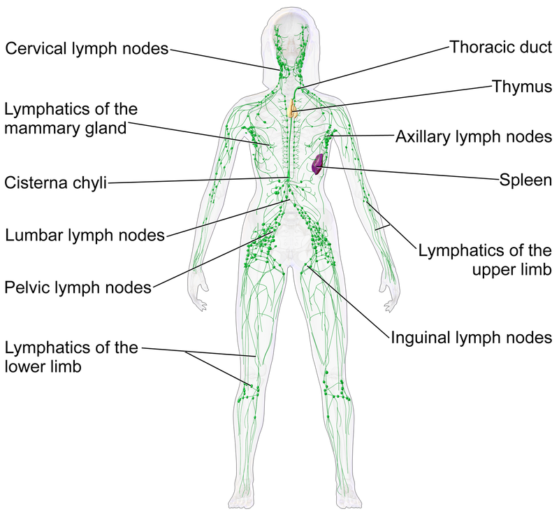
The lymphatic system
Blausen_0623_LymphaticSystem_Female (322K) Medical gallery of Blausen Medical 2014
Lymphatic vessels have slightly muscular walls and a system of valves which ensures that lymph keeps moving in the same direction. They converge onto a number of lymph nodes and the spleen and thymus.

Many cells of the immune system - lymphocytes - B cells and T cells - originate from or mature in the lymphatic system.

The lymphatic system is also involved in the intake of fats and oils from the diet. The central section of each villus in the ileum - called the lacteal - leads into lymphatic vessels which connect to the receptaculum or cisterna chyli in the top of the abdomen

Eventually lymph re-enters the blood system via the left subclavian vein in the armpit region.

Formation of tissue fluid

Ins and outs of capillaries

tissue_fluid (51K)

Tissue fluid provides what cells need, as well as removing wastes
The aorta carries (oxygenated) blood at pressure, although this pressure drops as it travels further from the heart and arteries branch off to supply each organ of the body. Pressure also drops as arterioles branch off to enter tissues within the organs, and these divide to supply a large number of capillaries.

Capillaries have a very thin wall, and hydrostatic pressure originally provided by the contraction of the left ventricle forces liquid out of the blood plasma. This liquid is mostly water with small ions (e.g. Na+, Cl-) dissolved in it, together with dissolved oxygen which is/was in equilibrium with oxyhaemoglobin in the red blood cells. It also contains dissolved glucose and amino acids, which are products of digestion and raw materials for building-up processes (anabolism) in cells. All these substances, which cells require, pass into the tissue fluid which bathes the cells of the organ.

Within capillaries, hydrostatic pressure falls from the arteriole end of the capillary to the venule end. This is because of loss of fluid and friction (against the lining of the capillary).

The fluid outside the capillaries has a lower hydrostatic pressure and the amount of fluid leaving the capillaries is proportional to the difference. Unlike the capillaries, there is not a marked pressure gradient in the tissue fluid outside.

Blood cells remain in the capillaries, and plasma proteins, e.g. albumin, do not cross the capillary wall, on account of their larger molecular size. As a result, a small (negative) water potential is maintained in the plasma so there is a tendency for water to re-enter the capillary by osmosis.

At the venule end, more water moves back into the capillary down the water potential gradient than is pushed out under the reduced hydrostatic pressure. Substances which cells need to excrete, e.g. carbon dioxide, are carried along in the tissue fluid and pass back into the blood. They eventually leave in the deoxygenated blood passing into the venule, and out into the vein, re-entering the circulatory system.


Putting water potential and pressure into context

Dissolved substances in the blood plasma give it a water potential which is of the order of -773 kPa. Most of this is caused by sodium and chloride ions which easily pass out of capillaries into tissue fluid, and thus they have an equal effect 'in each direction' so they can be ignored. However plasma proteins (especially albumin) which cannot leave the capillaries have a much lower osmotic effect resulting in a water potential of - 3.3 kPa. This is sometimes called the oncotic pressure or colloid osmotic pressure, and it tends to draw water back into the capillary.

In capillary beds the hydrostatic pressure varies according to position - higher at the arteriole end and lower at the venule end, but the water potential due to plasma proteins (albumin) retained in the blood in capillaries is constant. These two act in opposite directions.

In tissue fluid there is a lower but constant hydrostatic pressure, and also a constant but less negative water potential.

Putting numbers to the pressures and explaining the flow

arteriole_capillary_venule_figures (30K)

HP = hydrostatic pressure
WP = water potential due to plasma proteins

At A net pressure = > 1.2 kPa - working: >(4.3- 1.1) -(3.3-1.3) so liquid flows > out of capillary
At B net pressure = > -1.5 kPa - working: >(1.6-1.1)- (3.3-1.3) so liquid flows > into capillary

Other related topics on this site

(also accessible from the drop-down menu above)
Similar level
Exchanges with the environment
Surface area to volume ratio
Gas exchange
Digestion and absorption

Control of heart rate
Control of blood water potential
Control of blood glucose concentration

Levels of protein structure, exemplified by haemoglobin and myoglobin Full amino acid sequences of α- and β-chains
Simpler level
The heart and circulatory system

Interactive 3-D molecular graphic models on this site

(also accessible from the drop-down menu above)

Levels of protein structure shown by the haemoglobin molecule
Levels of protein structure shown by the myoglobin molecule

Web references


Gas exchange and diffusion in the book Regulation of Tissue Oxygenation.
Why is the partial oxygen pressure of human tissues a crucial parameter? Small molecules and hypoxia
The haemoglobin concentration of Chironomus cf.Plumosus l. (Diptera: Chironomidae) larvae from two lentic habitats
Blood proteins From Wikipedia, the free encyclopedia
Capillary Exchange - Chapter 20. The Cardiovascular System: Blood Vessels and Circulation
High blood pressure (hypertension) From theNHS

www.BioTopics.co.uk    Home     Contents     Contact via form     Contact via email     Howlers     Books     WWWlinks     Terms of use     Privacy