www.BioTopics.co.uk
Site author Richard Steane
The BioTopics website gives access to interactive resource material, developed to support the learning and teaching of Biology at a variety of levels.

Genetic diversity

This topic has connections with DNA, genes and chromosomes and DNA and protein synthesis (in Genetic Information). It continues with 'Genetic diversity and adaptation' . . . (links below)
Mouseover of green text (in the main body, not headings) should bring in further explanation in a small popup window.

Diversity and uniformity

Each species of organism has a number of shared features that are characteristic of that species. In general, these result from processes controlled by genes in the cells of the body.

In fact all the biochemical processes of the body depend on enzymes (which are proteins put together following genetic instructions from DNA), and it is easy to ignore the fact that most of the makeup of an organism is under genetic control. We humans usually have five fingers and five toes, and hair grows on our head (although perhaps not so reliably), and we don't grow feathers (but birds do!).

Observable features that are shared and common within a population have a basis in genes that are shared within a number of individual organisms. This is sometimes called a gene pool. Although this theoretically permits access to an exclusive variety of potentially shared genetic material, only a small fraction of it is practically accessible to any individual.

This unit explores the processes that produce and recombine the genetic diversity found in populations, whilst also supporting the reliable transmission of a proportion of inherited characteristics from one generation to the next.

Of course the process of mitosis also ensures that each individual usually has exactly the same genetic material in every cell of their body.


Amongst all living organisms there is a wide variety of easily observable features as well as those which can only be investigated at the biochemical level. Features which make organisms successful in life are likely to be carried over to the next generation when they breed, and this variation is the raw material for evolution.

Shared or common features are seen in groups of individuals called populations. When populations differ from other populations as a result of significant differences caused by inherited conditions they may be described as different species.

Gene mutations

A mutation is simply a change, and altering the sequence of bases in DNA means changing a gene. This spontaneous or random process may or may not result in changes to the protein or RNA produced by the cell, or it may be a change in a section of DNA that is not subject to transcription and translation. The most likely stage at which mutations occur is when DNA is replicating before cell division by mitosis or meiosis. During DNA replication, enzymes scan the resulting DNA strands and they may check the continuity of the molecular structure. Part of this process is called 'proofreading', and errors are usually efficiently dealt with, i.e. 'corrected' by another enzyme.

A mutation in an ordinary body cell is called a somatic mutation, and this may be passed on to other body cells produced by ordinary cell division involving mitosis. Some types of cancer are caused by mutation, and most result from a series of mutations.

The most significant mutations occur in cells that become gametes - germ line mutations. Following fertilisation, these can be passed on to the next generation, but they will only be noticed if they result in a change producing a dominant allele. Recessive alleles are much more likely to be masked by the other allele on the other (homologous) chromosome, which is provided by the (gamete from) the other parent. Over time, recessive alleles can spread through a population and be seen when two heterozygotes have children.

Gene mutations are fairly infrequent - sometimes described as 'one in a millon' events. However geneticists have produced mutant forms of many organisms through the use of mutagenic chemicals or radiation. See below


When a new version of a gene is produced, it is known as an allele. In many cases the new allele does not function as well as the original version, if it actually produces a product at all. It may be describd as a 'faulty gene'.

Often these alleles do not cause problems because in diploid organisms there are two sets of chromosomes, and every gene, which has a defined position on a specific chromosome (its locus) will have another version at the appropriate position on the partner chromosome. So if something is missing it may be made up for by the other allele. This is of course the background for dominance/recessiveness in inheritance.

For instance in many animals skin cells produce the brownish pigment melanin, which colours hair and skin to different extents. If this is not produced, the condition known as albinism occurs. It is caused by a recessive allele, so it must be inherited from both parents. In this case the cells do not produce the enzyme tyrosinase which is responsible for the production of the pigment melanin.

Science fiction and science facts

In some stories adult characters are subjected to radiation or chemical mutagens, and they are quickly transformed into another form.

But in real life mutation only affects a few cells, some of which may interact with other cells - fertilisation - and then the resulting zygotes take their time developing into adults.

Only if the allele is dominant will they eventually show the effect of the changed or mutant allele.

If the new mutant allele is recessive, individuals receiving the allele will not display any differences, and the allele will remain hidden in the population. Only after a few generations will the mutant alleles show their effect when two carriers (heterozygotes) reproduce together.

Mutagenic agents

Factors which increase the rate of gene mutations include certain chemicals, radiation and viruses.

Chemical agents that act as mutagens may damage nucleotides, or more specifically deoxynucleoside triphosphates (dNTPs), which are used by DNA polymerases to replicate DNA, or they can interfere with the cell's natural DNA repair mechanisms. Most mutagens are present in the cell under normal physiological conditions and they include reactive oxygen species (ROS) and alkylating agents.

Mutagens are also present in the environment and they include a multitude of chemicals that may be ingested in the foods that we eat, or in the air that we breathe. The radioactive gas radon is given off by certain rocks and may accumulate in some buildings.

Radiation such as ultraviolet (UV) light from sunlight or X-rays from electronic (including medical) equipment or gamma rays from radioactive atoms can be absorbed by the DNA molecule, causing it to vibrate at a molecular level, and if enough energy is passed on, bonds may break so that chromosomes split into pieces. Such pieces may not rejoin in their original configuration, so genetic material may be lost or reformed in a different sequence, or transferred to a different chromosome.

Mutagens that promote the development of cancer are called carcinogens.


Reactive oxygen species (ROS) include hydrogen peroxide, hydroxyl radicals and superoxides which are produced by cellular processes such as electron transport in mitochondria. There are several compounds that deactivate them, e.g. the enzyme catalase, but excess may sometimes escape this.
Of course, antioxidants are considered essential components of healthy diet, although they are not necessarily absorbed in the necessary amounts.
Oxidation of deoxyguanosine results in 8-oxo-2'-deoxyguanosine (8-oxodG) which causes a G to T transversion mutation.

Alkylating agents transfer methyl (-CH3) or ethyl (-C2H5) groups to nucleotide bases or the backbone phosphate groups. This may cause bases to mispair with inappropriate partners, e.g. (methyl) guanine may pair with thymine, instead of cytosine.
An example of an alkylating agent is ethylnitrosourea (ENU) which is known to induce point mutations, in particular A→T 'base transversions' Transversions involve changing a two-ring purine base (A or G) to a one-ring pyrimidine (T or C).
Used by geneticists as an experimental mutagen, ENU is known to cause fairly well defined mutations at an approximate rate of 1 per 700 gametes. It is apparently rather effective in targetting spermatogonial stem cells.

Many compounds have been found to cause cancer by this process (alkylation). For example, nitrosamines formed in foods by the action of nitrite preservatives.

And dangerous chemicals used in industry, e.g. vinyl chloride, and in warfare, e.g. 'mustard gas', have similar effects.

Base deletion

Consider a brief section of mRNA, which is produced by translation of a section of DNA. Here I have spaced out the bases into triplets and put the resulting amino acid codes to form a polypeptide below.
I have once again put my (simplified?) version of the genetic code opposite.
AUG ACA GGG AAA UCG GUC CGC CUU AAC CGC UGU UAG

met thr gly lys cys val arg leu asp arg cys STOP

Now imagine one of the DNA bases is deleted, and that really means removal of a whole nucleotide, and (following translation) this modification is transferred into mRNA. I decided to leave the first couple of triplets untouched, and put in an X to mark the spot of deletion.
AUG ACA XGG AAA UCG GUC CGC CUU AAC CGC UGU UAG
Converting into triplets again gives this:
AUG ACA GGA AAU CGG UCC GCC UUA  ACC GCU GUU AG?

met thr gly asp arg ser ala STOP thr ala val ?

Consequences of base deletions

You can see that most of the resulting amino acids are different, and in this particular case you see a stop codon which terminates the polypeptide chain and makes it shorter than before. A point mutation results in a 'frameshift', which changes the translation of mRNA into amino acids

It is highly likely that the resulting polypeptide will be significantly different at several levels of protein structure, and hence probably non-functional.

The random insertion of a nucleotide/base will have a similar effect.

Three deletions in a larger section of RNA might have a lesser effect, depending how far apart they are. In this case the STOP codon would cause chain termination at (almost) the right place.

The genetic code

A, C, G and U : 4 nucleotides/bases
N = any of them, R = purines (A or G) Y = pyrimidines (C or U)


RNA triplet codes amino acid abbreviation RNA triplet codes amino acid abbreviation
AAR lysine lys K GAR glutamic acid glu E
AAY asparagine asn N GAY aspartic acid asp D
ACN threonine thr T GCN alanine ala A
AGR
CGN
arginine arg R GGN glycine gly G
AGY
UCN
serine ser S GUN valine val V
AUR
(start)
methionine met M UAA UAG
UGA
(stop)
AUY isoleucine ile I UAY tyrosine tyr Y
CAR glutamine gln Q
CAY histidine his H UGY cysteine cys C
CCN proline pro P UGG tryptophan try W
CUN
UUR
leucine leu L UUY phenylalanine phe F

Base substitution

The swapping of one base for another is likely to have much less effect than missing a base. There is no frameshift effect, and the resulting polypeptide will be the same length (unless the base substitution is in the STOP codon).

Not all base substitutions cause a change

Because there are several - similar - codon possibilities for most amino acids (i.e. the genetic code is degenerate), there is the chance that single base sustitutions may result in the same amino acid as the unsubstituted base. This may be called a silent mutation. See the genetic code opposite.

This is especially true in the case of the third base in the codon triplet. Sometimes any other base (N) can act as a substitute, or else either G and C (R) or A and U (Y) are interchangeable.

Differences in the number of chromosomes

Changes in the number of chromosomes within cells can also count as mutations, but they do not cause a change in base sequence.

Problems with the separation of chromosomes in meiosis (chromosome non-disjunction) can result in gametes which do not have the normal (haploid) number of chromosomes, and after fertilisation this can result in a zygote which develops into offspring with different (mathematically odd) diploid numbers of chromosomes (in all the cells of the body, since they are produced following mitosis).

In humans there are several conditions involving an extra chromosome, so the body cells contain 47 chromosomes, not 46. This is caused by the incorporation of two copies of one of the chromosomes into a gamete, to be met with another single copy at fertilisation. This means that there are three copies of the affected chromosome - a condition known as trisomy. This can be seen when (pictures of) the chromosomes are arranged in order of size as a karyotype

It is also possible for a chromosome to be left behind when separation occurs in the production of gametes, so there may be only one copy in cells of an individual developing from the fertilised egg. This is called monosomy.

All of these instances of alterations to chromosome number cause development problems, and cardiovascular problems are quite common.


In humans there are a number of examples of conditions caused by an abnormal number of chromosomes. This is called aneuploidy.

Birth rate of Trisomy 21 in relation to maternal age Birth-rate-of-T21-in-relation-to-maternal-age-The-so-called-maternal-age-effect-was (21K)
Trisomy 21 - also known as Down('s) syndrome - results from an extra copy of the smallest chromosome. It is one of the most common chromosome abnormalities in humans, occurring in about 1 in 1,000 babies born each year. It is often caused by maternal non-disjunction, which is much more common with older mothers.

Pregnant mothers are generally offered a number of diagnostic test options as part of their antenatal care. In the past these have included invasive and somewhat risky techniques which extract cells of embryonic origin to be subjected to karyotyping in the lab:
chorionic villus sampling (CVS) which involves a biopsy of the placenta, and amniocentesis, in which cells are harvested from the amniotic fluid via a needle inserted into the amniotic sac.
More recent developments involve less problematic sampling of maternal blood in order to test for fetal DNA. Ultrasound scans, used for screening rather than diagnostic purposes, can give information about the development of the foetus, including potential heart defects and the neck region - 'nuchal translucency/folds' - which are characteristics of Down's syndrome.
Pregnancies confirmed with the diagnosis of trisomy 21 are often terminated via abortion. Those born with the condition have a number of health problems as well as reduced intellectual abilities - the brain typically has an excess of amyloid beta peptide produced, which is seen in Alzheimer's disease. Life expectancy has improved recently but a certain amount of assistance is necessary in later life.

Trisomy 18 (Edwards syndrome) and trisomy 13 (Patau Syndrome) present a number of developmental challenges. For each of these, the survival rate to 1 year old is only about 8%.

Turner syndrome (45,X, or 45,X0) is a monosomy due to the presence of only one X chromosome, and those affected are (infertile) females. It affects about 1 in 2000 baby girls. Unlike Down syndrome, this is not related to age of the mother, and it is probably caused by non-disjunction in the father.

Meiosis does more than halve chromosome number

Meiosis is a form of nuclear division but it is normally described in the context of the associated cell division. The cells produced - often known as daughter cells - are genetically different from one another, and the parent.

There are actually two nuclear divisions in meiosis, and as a result 4 haploid cells - each with n chromosomes - can be produced, although some may not be continued to maturity.
In particular, the production of female gametes often involves the discarding of a number of cells, or at least their chromosomes. Presumably this is in order to maintain a certain amount of cytoplasm, making the female zygote larger (and less able to move) whereas male gametes need to be more numerous and able to move.

As in mitosis, chromosomes move out of the nucleus and become attached to a spindle, to be moved to different ends of a cell before it divides. The same names are given to the stages in meiosis - prophase, metaphase, anaphase and telophase, but they are numbered I and II because of the two divisions.
Quite a lot happens during prophase I, and it may be subdivided into several sections.

Prophase I

me1 (9K)
me2 (8K)
Initially chromosomes condense so they appear to become shorter and thicker. In fact they have undergone DNA replication during the S-phase, and they consist of two chromatids, but this is more obvious at later stages.
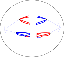
In these diagrams only 2 pairs of chromosomes are shown, and the colours denote maternal/paternal origin


me3 (9K) Each chromosome lines up with its homologous partner
Each chromosome becomes associated with its partner chromosome: in humans there are are 2 copies of chromosomes 1-22 which are described as homologous pairs, and of course they can be traced back to the two gametes which fused at fertilisation, so they can be described as being of paternal and maternal origin. They line up in pairs along their whole length, but X and Y are only similar for a short section so they only pair up in this region. These pairings are known as bivalents or tetrads. There are 4 strands alongside one another (each of which contains a double strand of DNA), held together by protein strands (the 'synaptonemal complex').
mex (8K)
Crossing over causes a chiasma (arrowed) to form

Since they are closely grouped, sometimes two of these strands may break and re-join, but with their partner strand. These are also known as non-sister homologous chromatids, and the mutual transfer of genetic material is known as crossing over. This means that the combination of genetic material (effectively the sequence of alleles) on these chromatids differs from the original chromatid. Genetic recombination has taken place. It is possible for this to take place at several points, producing more variation. Crossing over can also occur between sister chromatids, but the result is not significally different, unless unequal amounts of genetic information are exchanged.

As the homologous chromosomes separate, the points at which exchange of genetic material has taken place can be seen as cross-shapes where they overlap, and these chiasmata (singular: chiasma) gradually move to the ends of the chromosomes as they separate.

Chromosomes become attached to the spindle via the centromere region from which kinetochore fibres radiate.

Metaphase I

me5 (8K)
Chromosomes - each still consisting of a pair of chromatids - line up across the middle of the cell - the 'equator'.

Anaphase I

me6 (8K)
Each chromosome still consists of a pair of chromatids
Chromosomes move out along the spindle fibres towards opposite ends of the cell - opposite 'poles'. They appear to be pulled apart, and they have a characteristic V-shape.

Telophase I

A nuclear membrane forms around the chromosomes.

The cytoplasm divides, forming two cells, or it proceeds directly to the next stage, with the spindles typically at right angles to the previous ones.

Division II of meiosis

This is practically identical to mitosis, resulting in the separation of chromatids to separate nuclei inside four daughter cells.
mefinal4 (20K)
Each cell has only half the original number of chromosomes.
Crossing over has resulted in new combinations of alleles.

Meiosis results in genetic variation

In the production of sex cells, only half the genetic information of an ordinary body cell is passed on to the gametes: egg cells (ova) in females, sperm cells (spermatozoa) in males.

This may seem to ask a question: Which (genetic information) is kept and which is thrown away (or at least passed on to another cell)? The answer appears to involve several degrees of randomness.

In the first division of meiosis, homologous chromosomes are separated into two groups. But each group consists of a different combination of chromosomes. In humans, each resulting cell can have either the paternal or the maternal version of chromosomes 1-22 etc (as well as either one of the X chromosomes in females), and either a (paternal) Y or the (maternal) X chromosome in males.

The randomness of this separation into different groups means that the likelihood of daughter cells containing the same full set of chromosomes (and alleles) that they inherited from one of their parents is low.
In fact the possible number of different combinations of 23 chromosomes following meiosis is 223. This is 1 in 8,388,608.
This is ignoring the variation that may be introduced by crossing over.

You could be asked to suggest a formula using the expression 2n (also written as 2^n) (where n is the number of pairs of homologous chromosomes) to calculate such a number.

Random fertilisation introduces another degree of variation

Since both the male and female gametes show the same variation in combinations of chromosomes, the number of different combinations in the fertilised egg (zygote) are the product of the two i.e the square of this figure.

The possible number of different combinations of chromosomes following random fertilisation of two gametes described above (ignoring crossing over) would be 246, which is 7.037 ×10¹³.

Sources of variation in meiosis

The separation of homologous chromosomes in division I is called independent assortment or random segregation of chromosomes or genetic material. As the chromosome number is halved, the two sets of chromosomes that are dealt out contain one or the other of each homologue - which has its own, probably unique, combination of alleles.

Crossing over introduces another degree of difference between daughter cells, providing different base sequences and combinations of alleles between the two chromatids.

Separation of chromatids in division II results in the separation of similar nuclei, allowing the new combinations introduced by crossing over to be seen.

Life cycles

Sperm cell production
(spermatogenesis) sperm_production (22K) Human sperm cells are produced in the testes at the astonishing rate of 1500 per second, and the (maturing) process takes about 65 days!
How many of the resulting (4) sperms contain an X chromosome?
> 2 - half of them
Egg cell production .. plus (oogenesis) ..
egg_production (16K) Human egg cells are usually produced singly following division within follicles of the ovary, and they are the largest cells in the body. Some cells ('polar bodies') do not develop, and development of the main cell stops at certain stages.


In many organisms, meiosis is involved in the production of reproductive cells: gametes (sperm and eggs in animals, nuclei within pollen and ovules in flowering plants). These are haploid (n) - they have half the number of chromosomes of an ordinary cell which is diploid (2n).

In life-cycles there is usually a stage involving the combination of haploid cells (or at least their nuclei, containing their chromosomes). This is generally described as fertilisation, and it results in a diploid cell. Meiosis, involving halving of chromosome number, needs to alternate with fertilisation, which doubles it.

By combining the features of one organism with those of another, sexual reproduction provides raw material for evolution which is another way of looking at the next generation! And of course for the sake of equality only half of each parent's genetic material can be involved, and the selection of that half is subject to a shuffling process thanks to meiosis.

Both haploid cells and diploid cells can be increased in number by the process of mitosis. In the production of sperms, cell division by mitosis is a continuous process to provide cells which undergo meiosis.

In some organisms the diploid stage is the predominant one, and the haploid stage is temporary, but in other organisms the reverse is true.

Many plants show alternation of generations: They alternate between multicellular haploid sexual gametophyte stages and multicellular diploid asexual sporophyte stages.

Haploid spores produced by meiosis in the mature sporophyte germinate and grow into a haploid gametophyte, which grows and produces gametes by mitosis. Fusion of two gametes (syngamy) results in a diploid zygote.

This is the case in mosses and ferns. See below

The life cycle of a fern

This shows alternation of generations between the sporophye and the gametophyte stages Fern_life_cycle2copy (170K)

The gametophyte stage is also photosynthetic - it is just in blue to show it is haploid.
You should be able to able to name the processes occuring at the points marked A-G above.
A > meiosis B > mitosis C > mitosis D > mitosis
E > fertilisation F > mitosis G > mitosis

Which stage in the life-cycle requires the following environmental conditions?
dry/windy > spore dispersal
wetness > sperms swimming to meet egg

What is the significance of the sexual organs maturing at different times?
> It favours cross fertilisation - which increases genetic diversity

Flowering plants also show alternation of generations, but it is not so obvious, and there are several mitotic divisions accompanying the division by meiosis.

In the ovule inside the ovary of flowers, female megaspore mother cells undergo meiosis but only one of the four haploid cells continues to progress. In this respect it is similar to oogenesis in mammals, resulting in a large female gamete. The resulting megaspore nucleus divides three times by mitosis so the embryo sac contains 8 nuclei. One remaining at the edge near to the micropyle acts as a female gamete and will eventually fuse with a male nucleus coming down a pollen tube which enters at the micropyle, and two in the centre will also fuse with another male nucleus. The other nuclei appear to have guidance functions.

In regions of sporogenous tissue within the male anthers, microspore mother cells undergo meiosis, producing four haploid nuclei which enter separate pollen cells. Pollen itself is not a gamete but it contains haploid nuclei (produced by mitosis) and these function as male gametes. They are transferred to the female parts of the flower by pollinating insects or by being blown by the wind. One nucleus fuses with the female nucleus to form a diploid embryo and the other fuses with two female nuclei to form triploid tissue which develops into the seed's nutritive endosperm tissue. This is called double fertilisation. In fact these are preceded by another 'pollen tube nucleus' which controls the growth of the pollen tube from the stigma down the style and into the embryo sac.


Conversation between the attractive danseuse Isadora Duncan and Nobel prize winning poet, journalist, and novelist Anatole France, who were discussing eugenics, and the possibility of them producing a child together

Isadora (72K)
Anatole (20K)


Isadora: "Imagine a child with my beauty and your brains!"

Anatole: "Yes, but imagine a child with my beauty and your brains!"

OK both of these features are likely to be polygenic, i.e. caused by a number of genes/alleles so the child would probably be in the middle somewhere, but a few dominant alleles could swing things somewhat!

Comparison of meiosis with mitosis

Meiosis
Mitosis
Chromosome number Reduces (halves) the chromosome number (haploid cells from a diploid cell) Maintains the same chromosome number as in the parent nucleus
(Homologous) chromosome pairing? associate in pairs do not pair
Crossing over? +?Crossing-over & chiasmata formation No crossing-over
Number of divisions & offspring cellsTwo divisions → 4 offspring One division → 2 offspring
Resulting offspringGenetically different Genetically identical
An1 (17K)

Going through another stage

The diagram alongside shows a dividing cell from a human testis. Only four of the forty six chromosomes are shown inside the cell.

Which of the 4 stages of meiosis does this represent? Give a brief explanation why
> anaphase
> characteristic V-shape as chromosomes move from equator of cell towards poles
Which of the 2 divisions of meiosis does this represent? Give a brief explanation why
> I - (first division)
> each chromosome is seen to be double - sister chromatids have not separated
What term is used to describe the relationship between chromosomes circled at A and B?
> homologous chromosomes - although I have used the word 'partner'- and both would be given the same number
What term is used to describe the relationship between the structures B1 and B2 ?
> (sister) chromatids - copies of the same chromosome

The unlabelled structures on the right of the diagram show differently coloured sections.
What (genetic effect) does this indicate?
> recombination/reassortment of genes on one chromatid each
What (event within the cell) has caused it?
> crossing over - breaking and rejoining of sister chromatids in prophase I

Other related topics on this site

(also accessible from the drop-down menu above)

This series (genetic information)
DNA, genes and chromosomes
DNA and protein synthesis
Genetic diversity and adaptation

Species and taxonomy
Biodiversity within a community

(Cells and cellularity)
The cell cycle - especially mitosis

Web references

Rates of spontaneous mutation
Life Cycle of a Fern

But Suppose the Child Inherited My Beauty and Your Brains? - Thanks to Quote Investigator - I previously thought that George Bernard Shaw was involved.

Down syndrome From Wikipedia, the free encyclopedia

Survival of trisomy 18 (Edwards syndrome) and trisomy 13 (Patau Syndrome) in England and Wales: 2004-2011

Overview - Turner syndrome

www.BioTopics.co.uk    Home     Contents     Contact via form     Contact via email     Howlers     Books     WWWlinks     Terms of use     Privacy