|
Site author Richard Steane
|
The BioTopics website gives access to interactive resource material, developed to support the
learning and teaching of Biology at a variety of levels.
|
|
Each side consists of 2 chambers: the upper thin walled atria (singular - atrium) act as receiving chambers and the lower chambers, called ventricles, have thicker walls, because they actually do most of the pumping, by contraction.
Valves between the atria and the ventricles, and in the outflow from the ventricles, keep blood flowing in the correct direction.
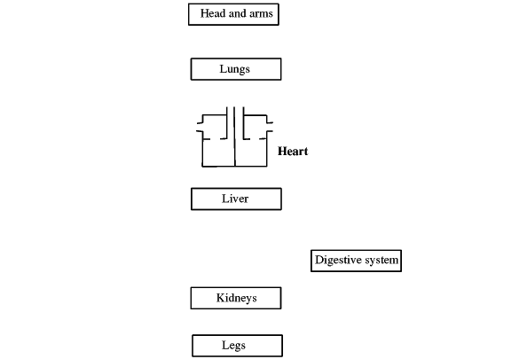
Use the mouse, or tap the screen, to label this diagram of a cross-section through the heart, and insert arrows to show the flow of blood:

Why are the parts on the left of the diagram labelled as right, and vice versa?
> inversion as in a mirror - looking at the position as in the body
Why is the wall of the left side of the heart somewhat thicker than on the right side?
> it has to work harder, to produce more pressure, to take blood all round the body, to the head and above
 What are the names of the 2 blood vessels running down over the outside of the heart?
>coronary artery >coronary vein
What does each of these blood vessels carry?
>oxygenated blood >deoxygenated blood
COMPLETE THE TABLE BELOW
NOTE: EACH IS A CONSEQUENCE OF THE ONE BEFORE
Types of Blood Vessel |
|||
|---|---|---|---|
| Notes | Arteries |
Capillaries |
Veins |
| blood direction | away from heart | between arteries and veins | towards heart |
| blood pressure | > high - nearest to heart | gradually runs from high to low | > low - "used up" |
| wall structure | > muscle and elastic layers | > single cell thickness | > muscle and elastic layers |
| wall thickness | > thick | > single cell thickness | > thin |
| internal diameter (also known as lumen, or bore) | >narrow; | >very narrow - down to size of red cell | > wider |
| valves present? | > no | > no | > yes |
| blood type, i.e state of oxygenation |
(usually) > oxygenated exception:pulmonary artery |
makeup of blood alters gradually, e.g. oxygen taken out or absorbed | (usually) > deoxygenated exception: pulmonary vein |
What causes (i.e. what gives the power for) the pulse you can feel at your wrist, for example?
> heart muscle contracting
What sort of thing is expanding when you feel the pulse?
> artery - not vein
The "Main artery" is called the aorta, and the "main vein" is called the vena cava.
Also label these blood vessels.

What do the different colours represent?
blue:> deoxygenated blood (blood with extra CO2)
red: >oxygenated blood
Blood enters and leaves most organs via a pair of blood vessels (artery and vein) which are named according to the organ.
What do you think is the name of the blood vessel bringing blood into the lungs?
>pulmonary artery
The hepatic portal vein is an exception, taking blood from the digestive system to the liver.
In what way do you expect that this blood may differ from other blood in the body?
>extra products of digestion (glucose, amino acids etc.)
Label the following on the diagram above: blood vessels leading to and from the lungs, liver, digestive system, and kidneys.